Project Description
Imaging techniques are used to diagnose metastases. In order to determine the exact stage of the disease, a biopsy can be carried out in which tissue samples are taken and histologically examined. A gentle and inexpensive imaging procedure is ultrasound examination. However, lymph nodes are often difficult to distinguish from the surrounding tissue in ultrasound images.
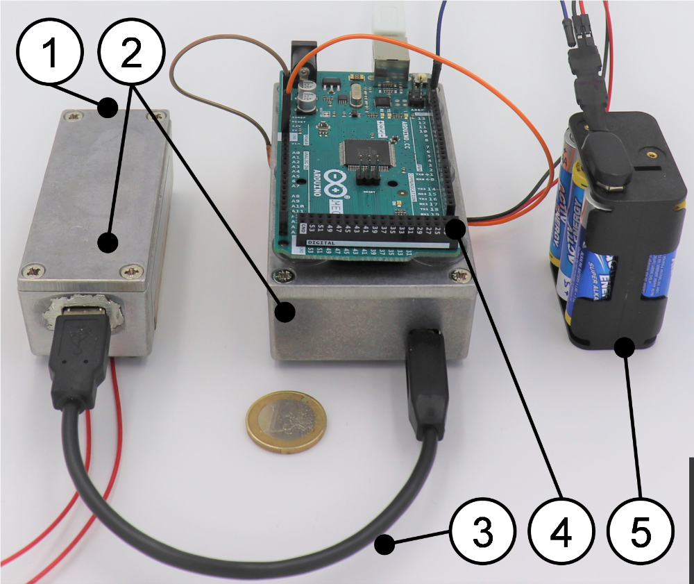
The Ultraclear research project is looking at the combination of ultrasound and scintigraphy to enable more precise detection of lymph nodes. To this end, a new type of device is being developed that combines an ultrasound probe and a specially designed scintigraphy module - comparable to a hand-held gamma camera - in one housing. The scintigraphy module uses specially arranged photodiodes to determine the three-dimensional position of a radioactively labeled lymph node. This position data is then fused with the ultrasound image. This procedure is intended to achieve a more precise identification of the lymph nodes and reduce the radiation exposure for the patient compared to conventional scintigraphic methods. The device is also said to be inexpensive to manufacture and use.
In addition, the removal of tissue samples should be made easier. A semi-autonomous biopsy guide is being developed for this purpose, which can be connected to the device. This guide enables automatic adjustment of the insertion angle based on the fused image data. This should both simplify the biopsy procedure and improve the visibility of the biopsy needle in the ultrasound image.
Overall, the project's research focuses on the following areas:
- Gamma detection
- Collimator design
- Detection algorithms
- Phantom production
- Image fusion
- Semi-autonomous biopsy
Publications
- Behling M, Wezel F, Pott PP, Miniature low-cost γ-radiation sensor for localization of radioactively marked lymph nodes, submitted